
Breast cancer remains one of the most prevalent forms of cancer affecting women globally. With significant advancements in medical technology, the tools for early detection and diagnosis have evolved. While mammograms are the gold standard for screening, the role of computed tomography (CT) scans in diagnosing breast cancer is often misunderstood. This article sheds light on the circumstances under which a CT scan may reveal signs of breast cancer and the importance of understanding its limitations and effectiveness.
Computed tomography, or CT scanning, is a medical imaging method that produces detailed cross-sectional images of the body using X-rays. This technology allows healthcare professionals to visualize various anatomical structures, including bones, organs, and soft tissues. CT scans are invaluable in diagnosing a variety of conditions, including trauma and diseases affecting internal organs. They create images that can be reconstructed into a three-dimensional view, providing a comprehensive look at the internal landscape of the body.
Typically, CT scans can be enhanced by using contrast agents, which may include iodine-based dyes. These agents improve the visibility of soft tissues and vascular structures in images, making it easier for physicians to identify any abnormalities. Despite their effectiveness in examining many internal structures, CT scans are not the primary tool for breast cancer detection.
While a CT scan can incidentally reveal signs suggestive of breast cancer, such instances are not the primary reason for its use. Studies indicate that up to 28% of CT scans conducted for other health-related concerns may inadvertently uncover suspicious breast lesions. For example, breast lesions may appear more distinctly on CT scans in certain situations, such as when they are located near the pectoralis muscle in individuals with dense breast tissue. However, these incidental findings should not lead to the assumption that CT scans are suitable substitutes for traditional breast cancer screening methods.
Generally, mammograms are the standard tool employed for breast cancer screening. They utilize low doses of X-rays to detect abnormalities in breast tissue and are associated with substantially lower radiation exposure compared to CT scans. In light of this, CT scans are best reserved for patients already diagnosed with breast cancer to evaluate tumor spread to other organs such as the lungs or liver.
Mammograms occupy a central place in breast cancer screening, especially for individuals who are asymptomatic. A routine screening can alert medical personnel to potential issues which may require further investigation through a diagnostic mammogram. If abnormalities are detected, healthcare providers may resort to a variety of follow-up imaging tests:
1. **Breast Ultrasound**: This imaging technique employs sound waves to create images, often used to differentiate between solid masses and cysts.
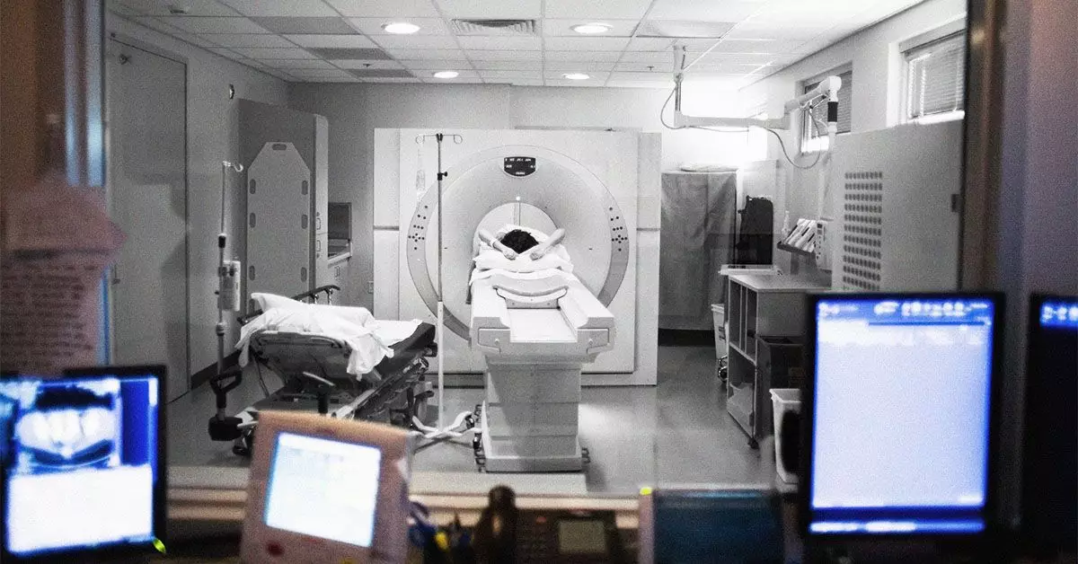
2. **Breast MRI**: Utilizing magnets and radio waves, MRIs offer a detailed view of breast tissue, making it an essential tool for complex cases or pre-surgical assessments.
3. **Biopsy**: In cases where imaging indicates suspicious areas, a biopsy enables the extraction of tissue samples for microscopic examination, definitive for cancer diagnosis.
In addition to these primary diagnostic methods, CT scans may be warranted for assessing breast cancer recurrence or spread, albeit as a supplementary approach rather than primary detection.
The efficacy of breast cancer treatment significantly improves with early detection. Regular screenings are vital even when there are no apparent symptoms. Individuals observing changes in their breasts—such as a lump or altered skin texture—should consult with healthcare providers promptly. Although these changes may sometimes indicate benign conditions, early evaluation is critical for achieving favorable outcomes.
Moreover, it is crucial to communicate any unusual breast changes effectively with healthcare providers, ensuring timely and appropriate interventions. Engaging in scheduled screenings is a vital step toward not only peace of mind but also potentially life-saving measures.
CT scans have a supplemental role in the diagnostic process for breast cancer, primarily indicated for assessing the extent of disease in diagnosed cases rather than being relied upon as a frontline screening tool. While incidental findings may provide helpful clues, they cannot replace the established modalities of mammography, ultrasound, or MRI. As awareness and technology advance, patients and practitioners alike must recognize the strengths and limitations of various imaging techniques in the fight against breast cancer. Making informed decisions about screenings and diagnostics is essential for promoting patient survival and health outcomes.