
Fundus photography is an innovative technique used to capture detailed images of the interior surface of the eye, particularly the retina. This method plays a critical role in identifying early signs of diabetic retinopathy, a serious eye condition that affects individuals diagnosed with diabetes. If left undiagnosed and untreated, diabetic retinopathy can lead to severe complications, including permanent vision loss and blindness. This article explores how fundus photography works, its relevance in diabetes management, and the impact of emerging technologies like smartphone imaging in enhancing eye care accessibility.
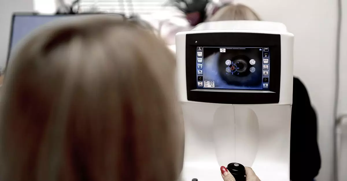
At the core of fundus photography lies the need for comprehensive eye examinations, particularly for those with diabetes. Fundoscopy, the procedure that involves using specialized cameras to photograph the fundus (the interior surface at the back of the eye), consists of capturing images that reveal critical information about the eye’s vascular health. These photographs can indicate various eye conditions, with diabetic retinopathy being one of the most concerning.
Diabetic retinopathy manifests through several specific changes in the retina, and fundus photography can unveil these alterations early on. Features such as microaneurysms—tiny lesions appearing as red dots—are among the first signs detected. Identifying these signs is crucial because timely intervention can significantly reduce the risk of severe vision impairment.
Several hallmark features may be visible in fundus images that denote diabetic retinopathy. Understanding these indicators can empower patients and healthcare providers to pursue timely diagnostic and therapeutic actions:
1. **Microaneurysms:** As previously mentioned, these small, round red dots are the earliest observable lesions in diabetic retinopathy. Their identification is vital for early diagnosis.
2. **Retinal Hemorrhages:** As diabetes compromises blood vessel integrity, ruptured capillaries can result in bleeding in the retina. These hemorrhages appear as dark, clearly defined patches and can manifest in various shapes.
3. **Hard Exudates:** These yellowish-white deposits signify lipid leakage from malfunctioning blood vessels, marking further progression of the condition.
4. **Cotton Wool Spots:** These fluffy white patches signify areas of reduced blood supply and are essential indicators of retinal stress.
5. **Neovascularization:** The formation of new blood vessels is a sign of progressive diabetic retinopathy. These vessels are often fragile and can lead to significant vision issues.
Understanding these characteristics emphasizes the importance of regular eye examinations for anyone with diabetes.
Recent advancements have broadened the scope of fundus photography, particularly through the advent of Selfie Fundus Imaging (SFI). This innovative technique allows individuals to capture fundus images using their smartphones after applying dilating eye drops. A 2022 study highlighting this method indicated that smartphone images taken by patients themselves were comparable in quality to images captured by trained technicians using traditional fundus cameras. This breakthrough holds promise for enhancing accessibility, allowing individuals to monitor their eye health conveniently and affordably.
However, while SFI demonstrates exciting potential, it must complement, not replace, professional eye evaluations. Proper training and understanding of the technique are essential to ensure high-quality imaging and accurate self-diagnosis.
For individuals with diabetes, early detection and management of diabetic retinopathy can significantly mitigate the risk of vision loss. Eye care specialists recommend comprehensive dilated eye exams at least once a year, with more frequent assessments for those with additional risk factors. Particularly for pregnant women or those with gestational diabetes, immediate eye evaluations are crucial to monitoring any rapid changes in eye health.
Symptoms of diabetic retinopathy, particularly in its early stages, may not be immediately apparent. As the condition progresses, individuals could experience fluctuating vision, floaters resembling cobwebs, or gradual difficulty in seeing detailed objects. Patients experiencing these or similar changes should promptly consult an eye care professional.
Fundus photography serves as a vital diagnostic tool in the detection and management of diabetic retinopathy. By regularly engaging in eye examinations and staying informed about personal eye health, patients can enact preventative measures against blindness related to diabetes. Moreover, integrating new technologies like SFI has the potential to enhance the accessibility of preventive eye care. Ultimately, the combination of vigilant monitoring, early diagnosis, and comprehensive treatment can prevent approximately 90% of blindness associated with diabetic retinopathy. Thus, individuals with diabetes must prioritize their eye health to safeguard their vision and overall well-being.